Understanding the Four Stages of Pressure Ulcers
Pressure ulcers, also known as bedsores or pressure injuries, occur when prolonged pressure on the skin restricts blood flow, leading to tissue damage. They often develop in individuals with limited mobility, such as those confined to a bed or wheelchair. Pressure ulcers progress through four stages, each requiring targeted treatment to prevent complications and promote healing. Advanced treatment options, including amniotic grafts, have shown promising results in accelerating wound healing, reducing inflammation, and supporting tissue regeneration. This blog will explore the stages of pressure ulcers, their symptoms, treatments, and how innovative solutions like amniotic grafts contribute to effective wound management.
What Causes Pressure Ulcers?
Pressure ulcers develop when prolonged pressure on the skin restricts blood flow, depriving tissues of oxygen and essential nutrients. The primary contributing factors include:
Prolonged Immobility
Individuals confined to a bed or wheelchair for extended periods are at a higher risk.
Friction and Shear
Repeated rubbing against bedding or clothing can weaken the skin, while shear occurs when the skin moves in one direction and the underlying tissues move in another.
Moisture
Excessive moisture from sweating, incontinence, or wound drainage can soften the skin and increase ulcer susceptibility.
Poor Nutrition
Malnutrition and dehydration weaken skin integrity and impair the body’s healing ability.
Medical Conditions
Conditions such as diabetes, peripheral artery disease, and circulatory issues increase the likelihood of developing pressure ulcers.
Now, let’s examine the four stages of pressure ulcers to better understand their progression and appropriate interventions.
Stage One: Early Skin Damage (Non-Blanchable Erythema)
Stage one pressure ulcers are the earliest form of tissue damage, presenting as persistent redness on intact skin. While the skin remains unbroken, underlying cells may already be compromised due to sustained pressure. This stage often serves as a warning sign that immediate action is needed to prevent further deterioration. Factors such as friction, shear, and moisture can exacerbate skin damage, making early intervention crucial.
Symptoms
Pressure ulcers in their early stages may present with subtle changes in the skin that can be easily overlooked. These symptoms often develop gradually and may become more pronounced with continued pressure or friction on the affected area. Recognizing these early signs is essential to prevent further progression and promote timely intervention:
- Non-Blanchable Erythema: Non-blanchable redness (erythema) that doesn’t fade with pressure release.
- Warm or Cool Skin: Warmth or coolness compared to surrounding skin, indicating changes in blood flow.
- Tenderness, Irritation, and Pain: One may experience tenderness, itching, or pain, which may be subtle but persistent in stage one.
- Firmness or Softness: Firmness or softness may appear in the affected area, suggesting early tissue inflammation.
Treatment Approaches
Effective intervention at this stage focuses on alleviating pressure and maintaining skin integrity:
- Pressure Relief: Regular repositioning to redistribute pressure and avoid prolonged contact with hard surfaces.
- Moisture Management: Use of barrier creams to protect against moisture damage from incontinence or perspiration.
- Skin Hydration: Maintaining hydration and using moisturizing agents to keep skin supple and resilient.
- Nutrition Support: Adequate protein, vitamins, and fluids intake strengthens skin and enhances its ability to recover.
Amniotic Grafts in Stage One
At this stage, direct application of amniotic grafts may not be necessary. However, amniotic-based topical treatments or skin creams enriched with amniotic-derived growth factors can enhance the skin’s healing potential by promoting cellular repair, maintaining hydration, and providing essential nutrients to support skin integrity.
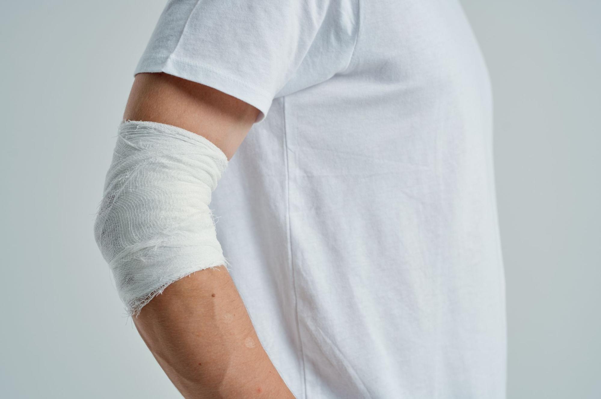
Stage Two: Partial Thickness Skin Loss
In stage two, the ulcer progresses beyond the epidermis, resulting in partial-thickness skin loss that exposes the dermis. The wound may appear as a shallow open sore or an intact/ruptured blister filled with clear fluid. This stage often signals an increased risk of infection, as the skin barrier is now compromised and more vulnerable to bacteria and irritants.
Symptoms
At this stage, visible changes in the skin may become more apparent, with the affected area showing signs of breakdown and increased sensitivity. These symptoms indicate a progression in tissue damage, requiring prompt attention to prevent further deterioration and potential complications:
- Pressure Relief: Regular repositioning to redistribute pressure and prevent prolonged contact with hard surfaces.
- Moisture Management: Use barrier creams to protect against moisture damage from incontinence or perspiration.
- Skin Hydration: Maintaining hydration and using moisturizing agents to keep skin supple and resilient.
- Nutrition Support: Adequate protein, vitamins, and fluids intake strengthens skin and enhances its ability to recover.
Treatment Approaches
Prompt treatment at this stage aims to support tissue repair and prevent further deterioration:
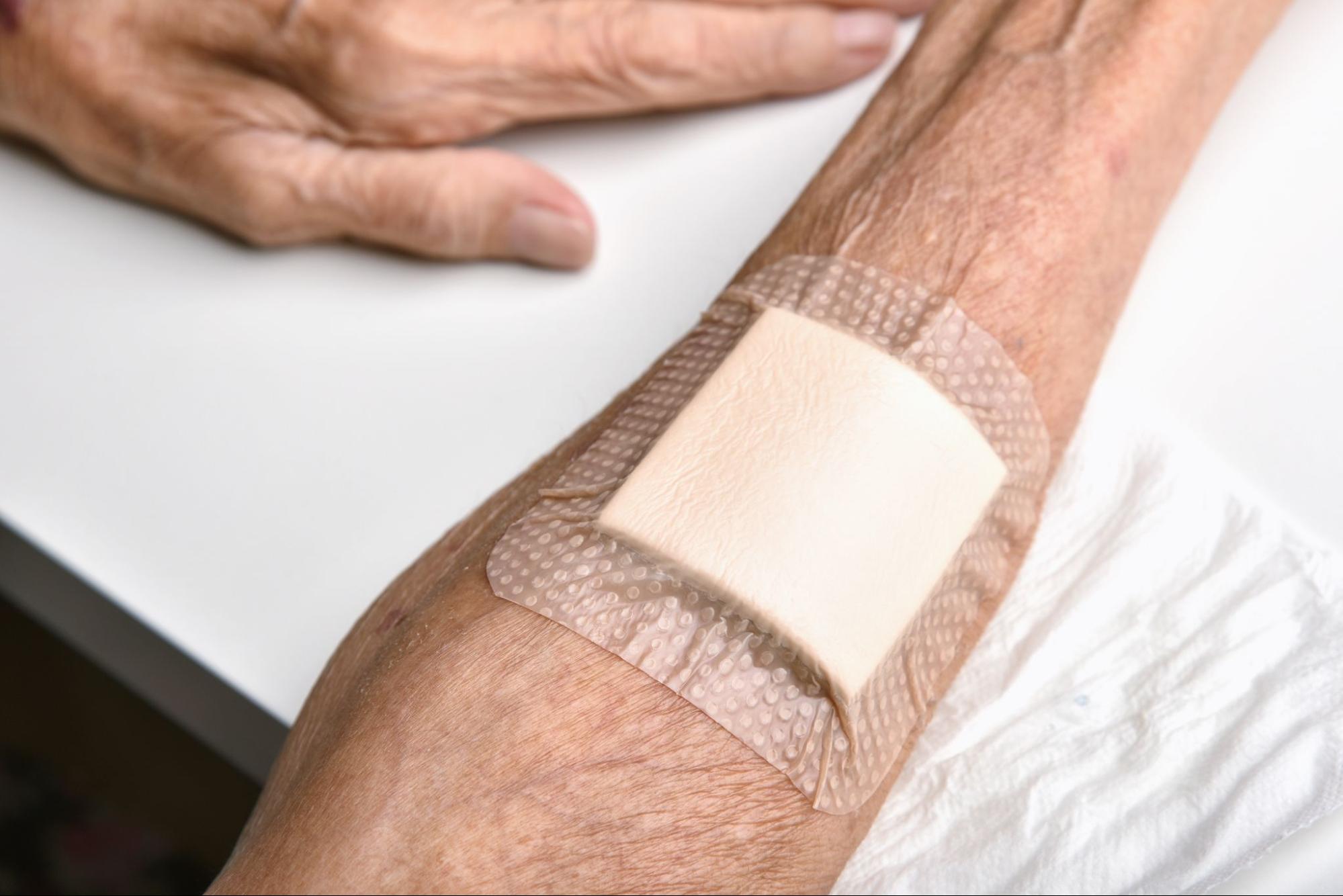
- Moist Wound Healing: Hydrocolloid or foam dressings that maintain an ideal healing environment by keeping the wound moist and protected.
- Debridement: Gentle removal of dead tissue to encourage new growth and prevent infection.
- Infection Prevention: Regular cleansing with saline or antiseptic solutions to remove bacteria and debris.
- Nutritional Optimization: Enhanced dietary intake, including zinc and vitamin C, is recommended to support tissue repair and immune function.
Amniotic Grafts in Stage Two
Amniotic grafts provide an excellent option for stage two ulcers by delivering growth factors, cytokines, and extracellular matrix proteins that accelerate tissue regeneration. These grafts can be applied directly to the wound to promote rapid epithelialization, reduce inflammation, and minimize scarring. Their natural anti-inflammatory properties also help alleviate pain and discomfort, supporting faster healing.
Stage Three: Full Thickness Skin Loss (Fat Exposure)
The ulcer extends deeper at stage three, affecting the subcutaneous fat tissue. The wound becomes crater-like, with the possibility of dead tissue (slough) and drainage. This stage significantly increases the risk of infection and complications as deeper tissues are exposed and healing becomes more complex.
Symptoms
At this stage, the wound may show deeper structural changes, with noticeable alterations in appearance and texture. These symptoms suggest a more advanced level of tissue damage that requires careful management to support healing and prevent further progression:
- Yellow or Necrotic Open Wound: One symptom is a deep open wound exposing fatty tissue, which may appear yellowish or necrotic.
- Presence of Slough: You may experience the presence of slough (yellow, necrotic tissue) that needs to be removed for healing to occur.
- Odor and Drainage: Foul odor and increased wound drainage, indicating possible infection.
- Red, Painful Ulcers: Pain and redness around the ulcer may worsen without proper intervention.
Treatment Approaches
Aggressive wound care is needed to promote healing and prevent infection:
- Debridement: Surgical, enzymatic, or autolytic removal of necrotic tissue to prevent further breakdown and allow healing.
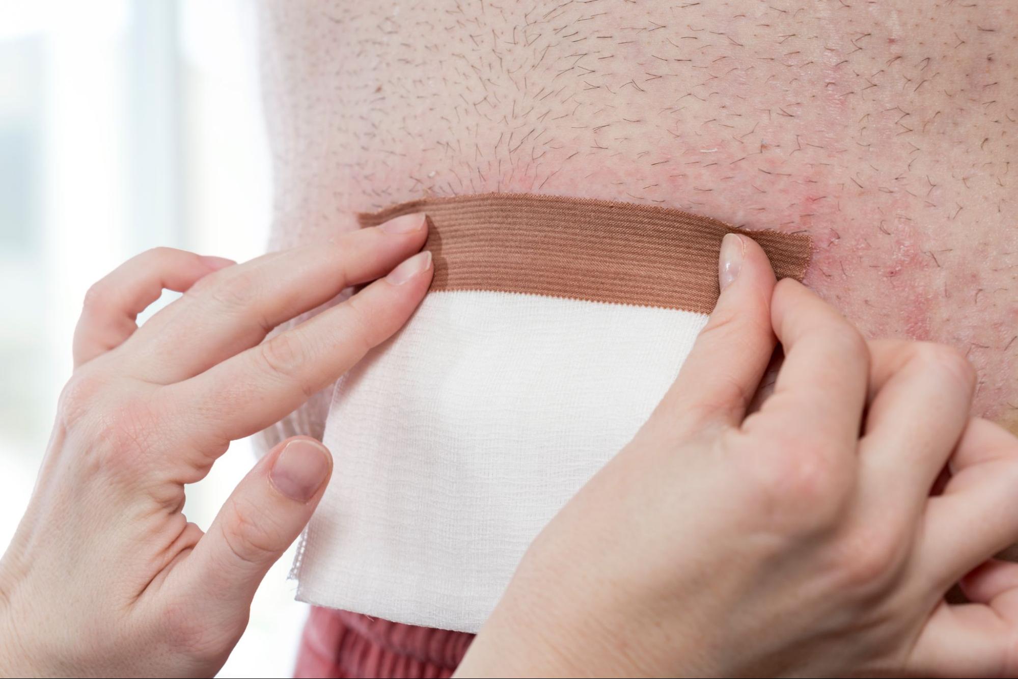
- Advanced Dressings: Alginate or hydrogel dressings are used to manage exudate, support healing, and keep the wound moist.
- Negative Pressure Wound Therapy (NPWT): Vacuum-assisted dressings that promote faster healing by enhancing blood flow and reducing bacterial load.
- Antibiotic Therapy: If signs of infection are present, oral or topical antibiotics are required to prevent systemic complications.
Amniotic Grafts in Stage Three
Amniotic grafts are highly beneficial at this stage, as they provide a protective scaffold that encourages the formation of granulation tissue, reduces pain, and speeds up the healing process. Their antimicrobial properties help reduce the risk of infection, making them an essential part of comprehensive wound care for deeper ulcers. These grafts also support collagen production, critical for wound closure and tissue strength.
Stage Four: Full Thickness Tissue Loss With Deep Exposure
Stage four pressure ulcers represent the most severe form, with full-thickness tissue loss exposing deeper structures such as muscles, tendons, and bones. The wound has a high risk of severe infection and complications such as osteomyelitis and sepsis. Healing at this stage is prolonged and often requires surgical intervention.
Symptoms
The affected area may show significant deterioration in advanced stages, with deeper tissues becoming visibly involved. These symptoms highlight critical damage and the urgent need for comprehensive care to address the injury and prevent further health risks:
- Deep Wound With Exposed Bone: A large, deep wound with exposed bone, tendon, or muscle is often accompanied by severe pain.
- Dead Tissue: Black or brown eschar (dead tissue) must be removed to allow healing.
- Nerve Damage: Severe pain or loss of sensation due to nerve damage from prolonged exposure.
- Wound Drainage: Extensive wound drainage with a strong odor, indicating deep tissue infection.
- Swollen, Red Tissue: Surrounding tissue may appear swollen and red, signaling systemic involvement.
Treatment Approaches
Stage four ulcers require immediate medical intervention and specialized care:
- Surgical Debridement: Extensive removal of necrotic tissue is often necessary to promote healing.
- Reconstructive Surgery: Skin grafts or flaps to close large wounds and restore function.
- Intravenous Antibiotics: Treat systemic infections and prevent sepsis, a life-threatening complication.
- Advanced Wound Care Techniques: To minimize pressure, biologic dressings, negative pressure therapy, and specialized beds are used.
Amniotic Grafts in Stage Four
Amniotic grafts provide a regenerative solution to aid tissue reconstruction and wound closure in severe wounds. They offer a biologically active matrix that stimulates cell growth, enhances collagen production, and accelerates the healing of deep tissue damage. Their anti-inflammatory properties also help manage wound pain and reduce the risk of infection, making them a crucial addition to advanced wound care strategies.
Benefits of Amniotic Grafts in Pressure Ulcer Management
Amniotic membrane grafts, derived from the placenta, are rich in growth factors, cytokines, and extracellular matrix components that contribute to wound healing in several ways. These biological components help create an optimal environment for cell migration, differentiation, and regeneration, making them a powerful tool in wound care. As a natural and effective solution, amniotic grafts provide a non-invasive option that complements standard wound care practices.
Accelerated Healing
This treatment enhances cell proliferation and tissue remodeling by providing essential nutrients and growth factors stimulating skin and tissue regeneration. Compared to traditional treatments, this results in faster wound closure and significantly reduced healing time.
Reduced Inflammation
Lowers inflammation and pain levels in chronic wounds by modulating the body’s immune response and promoting a more balanced healing environment. The anti-inflammatory properties help decrease swelling and discomfort, improving patient comfort and adherence to treatment.
Infection Control
Amniotic tissue provides a natural barrier against bacteria, reducing the risk of complications by preventing pathogens from penetrating the wound bed. The antimicrobial peptides within amniotic tissue further enhance its ability to combat infections and promote a sterile healing environment.
Minimal Scarring
This treatment promotes organized tissue regeneration with minimal fibrosis, helping the skin regain its strength and elasticity without excessive scar formation. This regeneration is particularly important for wounds in visible areas where cosmetic outcomes matter, ensuring a smoother healing process.
Biocompatibility
Amniotic grafts are naturally derived and well-tolerated by most patients, reducing the risk of allergic reactions or rejection. Since they are immunologically privileged, they do not trigger an immune response, making them suitable for individuals with sensitivities or chronic conditions.
Stages of Pressure Ulcers and the Role of Amniotic Grafts
Understanding the four stages of pressure ulcers is crucial for timely intervention and effective treatment. Whether at the earliest stage or the most severe, appropriate care can prevent complications and promote healing. Early recognition of pressure ulcers, combined with strategic management techniques, ensures better health outcomes and reduces the burden of long-term care. Innovative solutions such as amniotic grafts offer a groundbreaking approach to wound healing. They provide the necessary biological support for tissue regeneration and infection control. By leveraging the regenerative properties of these grafts, healthcare providers can improve healing rates and reduce complications associated with chronic wounds.
Visit the Stem Health Plus LLC blog for expert advice, innovative treatment solutions like amniotic grafts, and essential tips for effectively managing and preventing pressure ulcers. Stay proactive and empowered with the latest in wound care advancements!